
")


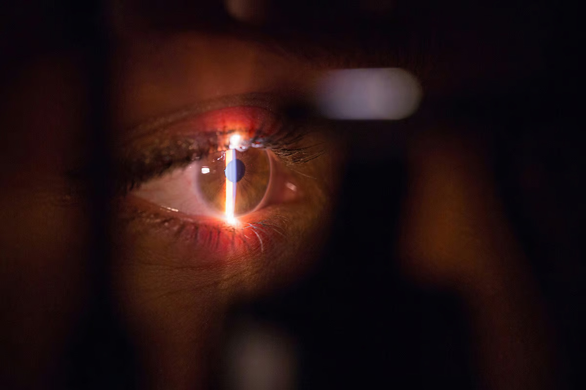 As science-fiction-y as it sounds, tooth-in-eye surgery has proven to be effective in dozens of cases over the last six decades. Brands&People / Unsplash
It’s called Osteo-odonto-keratoprosthesis (OOKP), and it’s actually not all that new. In fact, it was pioneered more than 60 years ago in Italy by ophthalmic surgeon Benedetto Strampelli. This procedure has been carried out dozens of times in a handful of countries over the last few decades, and Canada now has three patients who’ve undergone the first part of their two-stage OOKP surgery.
The first of these patients is 74-year-old Gail Lane, who lost her sight a decade prior. Her surgery was performed by Dr. Greg Moloney, an ophthalmologist at Providence Health Care in Vancouver who’s previously carried out the tooth-in-eye procedure on seven patients in his home country of Australia.
–
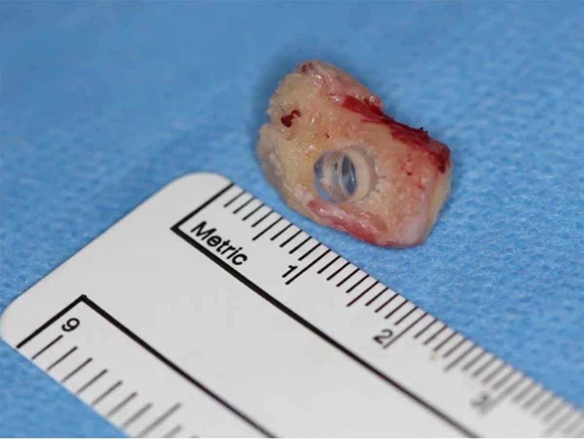
Here’s how it works: First, the patient’s canine tooth (also known as the eyetooth, due to its position directly under the eye) is extracted and shaped into a rectangle, and a hole is drilled into it to fit a plastic optical lens. Next, this tooth-lens is surgically embedded into the patient’s cheek for about three months, during which time a layer of tissue grows around it.
In addition, a patch of skin is taken from inside the patient’s cheek, and sewn onto the eye beneath the eyelid. When the tooth composite with its layer of tissue is ready, this flap of skin sewn on the eye is lifted, the damaged iris and lens are removed, and the tooth composite is inserted.
Finally, the flap of skin will be laid back over the eye to keep the tooth in place, with a hole cut in the flap to let light into the lens.
Patients typically start to regain vision a month after the surgery is complete.
Watch this animation below for an overview of the procedure.
–
[For the balance of this very interesting articles, please visit: https://newatlas.com/medical-tech/tooth-eye-surgery-ookp/]
Sources: Smithsonian Magazine, Vancouver Sun