")


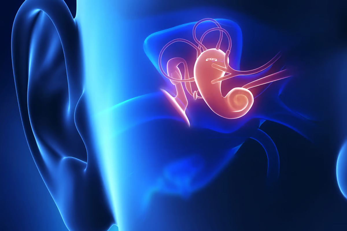 The discovery of a strange mechanism between the ear and the brain could lead to a new potential tinnitus treatment. Depositphotos –
Sensory hair cells are tiny structures in the cochlea that wave like blades of grass in the wind – but in this case, it’s the pressure of sound waves that gets them moving. When they do, they create electrical signals that are funneled through nerve fibers to the brain, to process what you’re hearing.
But a small percentage of these nerves actually run in the opposite direction, from the brain to the cochlea. Scientists have long been puzzled by the function of these backwards channels, and it’s hard to study their activity while people or animals are awake.
In the new study, scientists at the University of Southern California (USC) used an intriguing imaging tool to see what’s going on in there. The technique is called optical coherence tomography (OCT), which involves creating a 3D image of tissue using light waves. It’s currently used to scan the retina to diagnose conditions like glaucoma, but the team adapted it for use in the ear.
“OCT lets us look down the ear canal, through the eardrum and bone into the cochlea, and measure how it’s working – non-invasively and without pain,” said John Oghalai, lead author of the study. “What’s exciting about this is it lets us study how the brain is controlling the cochlea in real time.”
–
[For the balance of this very important article on Tinnitus please visit: https://newatlas.com/biology/tinnitus-treatment-blocking-back-channels-ear/]
–
The research was published in the Journal of Neuroscience.
Source: USC
–