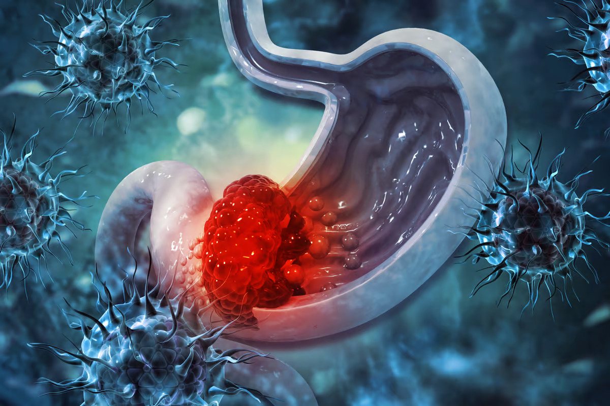
 A common, usually harmless bacteria caused stomach cancer in mice. Depositphotos –
About half of the world’s population carries the usually harmless bacteria Helicobacter pylori. While it’s well-known in the medical community that when H. pylori turns pathogenic, it can cause an infection that significantly increases the risk of gastric, or stomach, cancer, only 1% to 3% of those infected with the bacteria develop the condition.
The low percentage of people who develop gastric cancer because of H. pylori infection has led scientists to question whether there’s an additional pathogen contributing to the disease. It prompted researchers from the Nanyang Technological University (NTU) Singapore and the Chinese University of Hong Kong (CUHK) to co-lead a study investigating what that microbe could be.
The researchers examined the non-H. pylori gut microbiome in patients with different stages of gastric cancer, from superficial gastritis (inflamed stomach lining) to atrophic gastritis (thinning of the stomach lining and loss of cells that release substances to aid digestion) to intestinal metaplasia (stomach lining cells are replaced by cells similar to intestinal cells), and finally to cancer. They found five oral pathogens enriched in these patients’ gastric linings, including Streptococcus anginosus.
S. anginosus is part of the normal flora of the mouth, nose, throat, gut, and vagina. It usually doesn’t pose a problem for healthy people but can cause opportunistic infections when the body’s immune system is weakened. The bacteria’s role in gastric cancer, including its mechanism of action, has remained largely unclear.
Using mouse models, the researchers found that colonization with S. anginosus initiated an acute inflammatory response followed by a chronic phase with intensive and persistent gastritis. Chronic inflammation is known to trigger cancer growth and development. In the mice, infection caused a progression from chronic gastritis to atrophy, metaplasia, and dysplasia, the same pathway that humans follow before they develop gastric cancer. Further, co-infection with S. anginosus and H. pylori produced greater gastric inflammation than either pathogen alone, suggesting that the two might act together to promote gastritis.
Introducing S. anginosus into germ-free mice – mice born without microorganisms in or on them – produced the same precancerous changes. This implied that S. anginosus alone, but not its interaction with the gastric microbiome, was enough to drive cancer formation.
“H. pylori infection and family history are widely recognized as the two main risk factors for gastric cancer,” said Joseph Sung Jao Yiu, co-corresponding author of the study and Emeritus Professor of Medicine at CUHK. “The discovery of enrichment of S. anginosus in the gastric mucosa across different stages of cancer opens up a whole new direction in understanding the pathogenesis of gastric cancer. We also wish to flag that co-infection of H. pylori and S. anginosus leads to an even higher risk of precancerous atrophy, metaplasia, or gastric cancer.”
The researchers examined the mechanism underlying this process and found that S. anginosus used its surface protein, TMPC, to communicate with the Annexin A2 (ANXA2) receptor on cells in the stomach lining. The interaction allowed the bacteria to attach to and colonize the cell, activating mitogen-activated protein kinase (MAPK), an enzyme that coordinates cell proliferation, differentiation, and survival. Knocking out the ANXA2 receptor stopped S. anginosus from activating MAPK and contributing to cancer progression.
“We have established the role of S. anginosus in gastric carcinogenesis and its related mechanism; next, we will explore the therapeutic potential of targeting it to reduce gastric inflammation and cancer risk,” said Yu Jun, director of the Institute of Digestive Disease at CUHK and one of the study’s corresponding authors.
The study was published in the journal Cell. Source: NTU Singapore, CUHK
–


